


The organization of Drosophila wing epithelial cells after wing inflation. Iyengar, Balaji (2012). figshare. License CC-BY
As I child I wondered what would happen if my bones grew at a different speed to my skin? Would the bones pop out or would I have floppy boneless regions? How did everything coordinate? As with most childhood questions, below the surface of the apparently childish simplicity there is a deep scientific question which parents often battle to answer.
Your body’s fantastic ability to self-assemble and self-organize so that body-wide function and cooperation emerges from the small-scale actions of individual cells is just one of the wonders of the living world.
This work of coordinated art is an active area of research. The level of complexity is amplified by the variety of different ‘players’, interactions, feedback controls and chemical signals which vary over time and space. Within this diverse system we will zoom in on one tissue type.
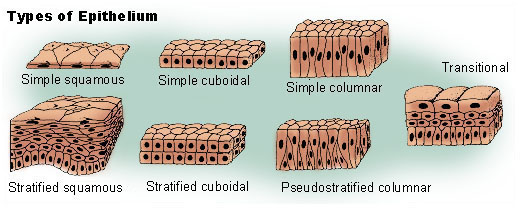
“Illu epithelium”. Licensed under Public Domain via Wikimedia Commons
Epithelial tissue is made of sheets of cells which line body surfaces, cavities and organs, and these cells are the most prolific type of cell in human bodies. Epithelial cells maintain a closed barrier to the external environment and so protect the system they line, alongside a variety of other location-dependent functions. However, scientists studying the epithelial layer have found that the large-scale function of this layer is in fact created and maintained by a surprisingly simple mechanism acted out on a small scale by many simple individuals. This link between local and global – how local cell action results in high-level tissue structure – is important in learning more about these cooperating, self-organizing systems.
![Drosophila melanogaster By André Karwath aka Aka (Own work) [CC BY-SA 2.5], via Wikimedia Commons](https://www.faraday.cam.ac.uk/wp-content/uploads/2020/02/drosophila_melanogaster_-_top_aka.jpg)
Drosophila melanogaster By André Karwath aka Aka (Own work) [CC BY-SA 2.5], via Wikimedia Commons
Upon closer inspection it has been found that the epithelial cells in this layer are different shapes, with 4-9 sides. The impression this creates is one of a cobblestoned street as opposed to the more uniformly structured regularity of a honeycomb. If tasked with building a wall, would you not use same-sized bricks? Similarly, would you not expect an ordered system to be based on consistent shapes? Further research into the different shaped cells shows that they are consistently found in specific ratios. Not only that, but these same ratios are observed across different animal groups. How does this irregular and yet consistent layout come about through the behaviour of individual cells?
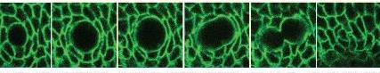
Cell Division Captured by Fluorescent Marking, non-exclusive license from Nature Publishing Group
Using laboratory techniques, Gibson and his colleagues were able to make the cell interfaces fluoresce so they could make time-lapse movies of the growth dynamics. They saw no movement of cells to suggest that they were being sorted into a specific arrangement. Instead, simple mitosis (cell division) is enough to explain the emerging pattern.
The team of scientists modelled this process using mathematics and computer models to predict, based on rules of cell biology and mitosis, an equilibrium of approximately 29% petagons, 46% hexagons, 21% heptagons and lesser frequencies of other polygons. They then painstakingly counted the different types of epithelial cell shapes in real specimens and confirmed that the predicted ratios were indeed reflected in all the organisms they checked; frogs, freshwater hydra and fruit flies. So although the epithelial landscape appears irregular, it is not random – order emerges[1].
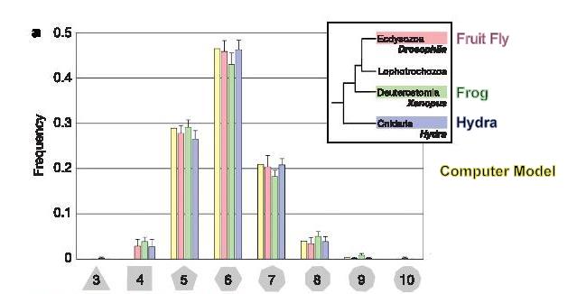
Cell Shape Counts, non-exclusive license from Nature Publishing Group
The question remains, how can cell division cause this diversity of shapes but maintain consistency in the proportions of different shapes? In the epithelium, cells stick to their neighbours. Cell division changes the number of neighbours, which changes the number of sides to the cell. This process will happen repeatedly, so the number of sides a cell has will fluctuate during development.
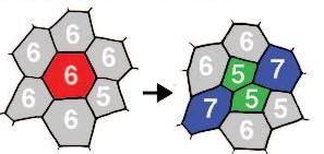
Cell Division, non-exclusive license from Nature Publishing Group
Cells gain additional sides as their neighbours divide. When it is their turn to divide, the daughter cells have fewer sides. Amazingly, these gains and losses in the number of sides balance out, resulting in the shape distribution discovered.
The principles of cell adhesion and mitosis are fundamental to biology, so this simple mechanism can be used to study a variety of similar systems across many different animal groups. Furthermore, the study of self-organising biological systems can also bring insights into the fields of robotics and computer science.
The scientists who work on this project collaborate with Harvard University’s ‘Self-organizing Systems Research Group’ who state the following…
“Biological systems, from multicellular organisms to social insects (“superorganisms”), get tremendous mileage from the cooperation of vast numbers of cheap, unreliable, and limited individuals. As we build artificial systems with similar characteristics — robot swarms, modular robots, sensor networks, programmable materials — can we achieve the kind of complexity and reliability that nature achieves?”
This tissue’s ability to generate and maintain order during growth is one of the many wonders of developing bodies. However, with it we are just scratching the surface of the myriad of processes which run, unnoticed by us, to organise and shape our bodies.
References:
- Matt Gibson, Ankit Patel, Radhika Nagpal, Norbert Perrimon (2006). The Emergence of Geometric Order in Proliferating Metazoan Epithelia, Nature, 442(7106):1038-41. (nature link) , (pdf), (supplement)
- William Tyler Gibson, J. Veldhuis, B. Rubinstein, H. Cartwright, N. Perrimon, W. Brodland, Radhika Nagpal, and Matthew C. Gibson (2011). Control of the Mitotic Cleavage Plane by Local Epithelial Topology, Cell Volume 144, Issue 3, 414-426, (pdf), featured in Cell highlights (” Funky shapes and Pushy neighbors”)
Nature Group Images:
Reprinted by permission from Macmillan Publishers Ltd: [Nature] (Reference (1) above), copyright 2006
 Cara Daneel is a Research Assistant supporting the work of Dr Ruth Bancewicz at The Faraday Institute. After receiving her Marine Biology and Oceanography degree from the University of Cape Town, South Africa, Cara has worked in conservation and education in a variety of countries; both in the Northern and Southern Hemispheres.

Explore more
resources for churches:
Authors
Subscribe to Faraday Churches
Get email notifications as new resources are published: